As dental professionals, we are often faced with complex cases where the standard diagnostic tools and techniques don’t provide all the answers we need. In these situations, Cone Beam Computed Tomography (CBCT) has emerged as a game-changer. For trained dentists, CBCT offers a level of precision and insight that significantly improves treatment planning and clinical outcomes, particularly in challenging dental implant cases.
In this blog, we’ll dive into several real-world case studies where CBCT imaging played a pivotal role in diagnosing issues, planning procedures, and ultimately improving patient outcomes in complex implant cases.
1. Case Study 1: Implant Placement in the Posterior Maxilla with Sinus Proximity
Challenge:
A 55-year-old female patient presented with a missing upper molar and a history of sinus issues. The patient had been wearing a partial denture for several years but wanted a permanent solution through dental implants. However, the posterior maxilla in the area of interest showed limited bone height, and traditional 2D X-rays did not provide adequate details on the relationship between the sinus cavity and the available bone.
Solution with CBCT:
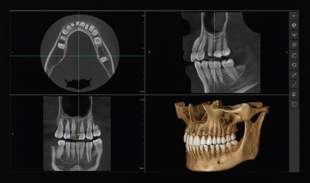
Using CBCT, we obtained a 3D view of the patient’s maxillofacial structures. The images revealed not only the bone density and volume but also the exact proximity of the maxillary sinus. The CBCT scan highlighted areas of atrophy in the bone, but more importantly, it identified a small pocket of bone that could be utilized for implant placement, which wasn’t visible on conventional radiographs.
Outcome:
With the precise data from the CBCT, we were able to plan for a sinus lift procedure to augment the bone and prepare the site for implant placement. The 3D imaging also allowed for the selection of the ideal implant size and angulation to avoid sinus perforation. The result was a successful implant placement with optimal bone integration and no complications. The patient reported a smooth recovery and a significantly improved quality of life.
Key Takeaway:
CBCT was instrumental in visualizing the complex relationship between the bone and sinus cavity, which ensured a safer, more accurate treatment plan and led to a successful outcome.
2. Case Study 2: Implant Placement in a Narrow Mandibular Ridge
Challenge:
A 62-year-old male patient sought an implant solution for a missing lower first molar. Upon clinical examination and 2D X-rays, we discovered that the mandibular ridge was severely narrow due to past resorption. The available bone width was insufficient for a standard implant, and there was a concern about damaging the inferior alveolar nerve, which ran close to the implant site.
Solution with CBCT:
The CBCT scan provided a 3D cross-section of the mandibular bone and allowed us to measure the exact width of the alveolar ridge at multiple levels. The scan revealed that while the ridge was narrow, there was adequate bone available on the buccal and lingual aspects of the ridge to place a tapered implant at a slight angle. The CBCT images also gave us a clear view of the position of the inferior alveolar nerve, ensuring it was safely avoided.
Outcome:
Based on the detailed CBCT data, we proceeded with an angled implant placement. The results were excellent, with the implant integrating well into the bone, and the patient was able to return to normal function without complications. The precise information provided by CBCT allowed for a customized treatment plan that would have been difficult, if not impossible, to devise with traditional 2D imaging.
Key Takeaway:
CBCT’s ability to provide precise measurements of bone width and positioning of critical anatomical structures allowed us to create a treatment plan that was both safe and effective, improving the overall outcome of this challenging case.
3. Case Study 3: Full-Arch Implant Reconstruction with Severe Bone Loss
Challenge:
A 70-year-old male patient was referred for a full-arch implant reconstruction due to severe bone loss in both the upper and lower jaws. The patient had suffered from advanced periodontal disease, which resulted in significant atrophy of the alveolar ridge. Traditional X-rays failed to provide a comprehensive understanding of the extent of bone loss, and we needed a clear 3D representation of the remaining bone structure to plan for full-arch implants.
Solution with CBCT:
CBCT allowed us to view the entire edentulous area in 3D, not just in the horizontal plane but also in the vertical dimension. The scan revealed areas of bone grafting potential and areas where the bone was too thin to support conventional implants. It also highlighted critical structures, such as the maxillary sinus, inferior alveolar nerve, and mental foramina, that would influence implant placement.
Based on the 3D data, we were able to identify areas where bone grafting was necessary and plan for zygomatic implants in the maxilla to provide the needed support. In the mandible, we selected a combination of traditional and short implants to maximize bone use and minimize surgical trauma.
Outcome:
With the CBCT images guiding the surgical approach, we were able to restore both arches with implants successfully. The patient experienced a smooth recovery, and the prosthetic outcomes were highly functional and aesthetic. The ability to visualize the full bone structure and surrounding anatomy in 3D was critical to avoiding complications and achieving the desired results.
Key Takeaway:
For complex cases like full-arch reconstructions, CBCT provides a comprehensive view of the available bone and surrounding structures, allowing for more accurate planning, safer surgeries, and better long-term results.
4. Case Study 4: Implant Placement After Tooth Extraction with Limited Immediate Bone
Challenge:
A 45-year-old female patient required an implant following the extraction of a failing premolar. However, upon initial examination, we found that there was limited bone remaining after the extraction, which posed a challenge for immediate implant placement.
Solution with CBCT:
CBCT allowed us to assess the bone volume and density in the extraction site in 3D. The scan revealed that there was adequate bone on the buccal aspect of the ridge but a significant gap between the available bone and the implant site. Using the 3D data, we planned for a simultaneous bone grafting procedure with the implant placement, utilizing a guided surgery approach. The CBCT scan also enabled us to determine the optimal implant size and placement angle to maximize stability.
Outcome:
The combination of grafting and implant placement went smoothly, and the patient healed well with excellent bone integration. The 3D planning allowed us to deliver a precise implant placement with minimal post-operative complications. The use of CBCT in this case led to a more predictable and successful outcome compared to a traditional 2D approach.
Key Takeaway:
CBCT provides critical insights for cases where immediate implant placement is considered, particularly when bone volume is limited. It allows for precise grafting and implant placement, leading to more predictable and favorable outcomes.
Conclusion: The Power of CBCT in Complex Implantology
These case studies illustrate just how transformative CBCT can be in complex implant cases. By providing high-resolution 3D imaging of the bone, surrounding structures, and anatomical variations, CBCT enables dentists to plan more accurately, avoid potential complications, and deliver superior outcomes for patients.
For trained dentists, integrating CBCT into your practice isn’t just about having advanced technology at your disposal; it’s about improving your diagnostic capabilities and treatment precision. Whether you’re dealing with sinus issues, narrow ridges, bone loss, or immediate implant placements, CBCT can help you navigate the challenges of modern implantology with confidence and competence.
If you haven’t yet embraced CBCT in your practice, now is the time to consider its many advantages. The more precise your planning, the more successful your outcomes will be—and ultimately, your patients will thank you for it.