Providing the highest level of care requires choosing the right diagnostic tools for each clinical situation. While 2D X-rays remain fundamental in dental diagnostics, Cone Beam Computed Tomography (CBCT) has transformed how we approach complex cases in implantology, endodontics, orthodontics, and surgical planning.
This article compares traditional imaging and CBCT, outlining when to use each for optimal patient care.
Traditional Imaging: The Foundation of Dental Diagnosis
Pros:
- Widely Available: Found in nearly every dental office, suitable for routine care
- Quick and Cost-Effective: Fast image capture and minimal operational costs
- Low Radiation: Especially panoramic X-rays, making them safe for repeated use
Cons:
- Limited Detail: Offers only 2D views, with limited insight into depth or spatial relationships
- Overlapping Structures: Can lead to unclear images or misinterpretations
- Not Ideal for Complex Cases: Lacks detail needed for precise implant planning or surgical assessment
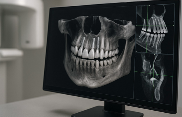
CBCT: A 3D Revolution in Dental Imaging
Pros:
- High-Resolution 3D Views: Visualize bone, nerves, sinuses, and soft tissues in full volume
- Improved Diagnostic Accuracy: Eliminates superimpositions and reveals hidden pathologies
- Enhanced Treatment Planning: Ideal for implant positioning, jaw assessments, and surgical simulation
- Guided Surgery Support: Enables custom surgical guide creation for millimeter-level precision
- Post-Operative Monitoring: Follow healing and detect complications with detailed visualization
Cons:
- Higher Radiation: Still relatively low, but greater than a single 2D radiograph
- Greater Cost and Space Needs: Requires investment in equipment, space, and training
- Not Always Necessary: May be excessive for routine procedures or low-risk cases
When to Use Each Modality
Traditional Imaging Is Best For:
- Routine checkups and cavity detection
- Basic orthodontic assessments
- Monitoring simple post-op healing
- Low-risk cases with no surgical intervention
CBCT Is Indispensable For:
- Dental Implants: Assess bone structure, density, and proximity to vital structures
- Complex Surgeries: Orthognathic surgery, third molar extractions, sinus lifts
- Endodontics: Root fractures, accessory canals, persistent infection
- Advanced Orthodontics: Impacted teeth, skeletal relationships, airway evaluation
- Pathology Diagnosis: Cysts, tumors, or unusual anatomical findings
Conclusion: Choose Based on Complexity and Diagnostic Need
Traditional X-rays remain essential for general dentistry, offering affordability and simplicity. However, when the stakes are higher—when precision, depth, and risk avoidance are key—CBCT should be your tool of choice.
The ideal approach?
Build your imaging protocol around patient-specific needs, using 2D imaging for routine evaluations and reserving CBCT for cases where 3D insight is essential. Integrating both smartly ensures clinical excellence, reduces complications, and elevates your diagnostic and treatment planning capabilities.